Background
Glenoid version measurement is critical in shoulder arthroplasty especially for the anatomic design in which the glenoid implant remains the weak link and where incorrect positioning leads to a high failure rate.1–3
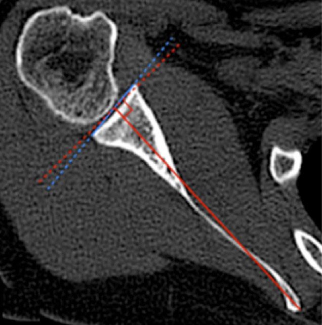
Essential knowledge of the glenoid vault anatomy and version is crucial for long-term survivorship. Many preoperative radiological methods have been proposed to calculate glenoid version, mostly based on CT scan using simple axial slices, 3D-corrected slices or even direct 3D viewing of the glenoid.4–7 The most commonly used method is the traditional Friedman’s angle4 but several limitations have been shown, notably the fact that it is dependant on the shape of the scapula body 6,8 and that the medial border of the scapula must be included in the CT scan field, which is not often the case.6 Moreover, glenoid version does not necessarily correspond to the orientation of the glenoid vault, a conical structure of cancellous bone that is sought for maximum purchase of pegs, keels or screws when resurfacing the glenoid. Matsumura et al. have recently proposed a new method of evaluating glenoid version according to the medial tip of the glenoid vault (the vault method), which they has shown to be more reliable than Friedman’s angle.6


